U-SPECT7CT from MILabs (a Rigaku company) offers unmatched preclinical dynamic and whole body Single Photon Emission Computed Tomography (SPECT) performance, integrated with diagnostic micro-CT.
With best-in-class spatial resolution of 0.25 mm for in vivo imaging, 0.12 mm for ex vivo specimens and high temporal resolutions of < 1 second for focused imaging as well as < 8 seconds for whole-body mouse imaging, U-SPECT7CT is ideal for your preclinical research projects, high-energy alpha & beta theranostics imaging and dosimetry. With the ability to upgrade to include other imaging modalities and to be used as PET/SPECT/CT and even PET/SPECT/OI/CT, U-SPECT7CT can grow with your needs.
Key features
SPECT module
- Ultra-high SPECT resolution - 0.25 mm in vivo, 0.12 mm ex vivo
- High sensitivity - up to 90,000 cps/MBq (when upgraded)
- High speed imaging – suits cardiac-, respiratory-, or dual-gated imaging
- Beds/cells for mice, rats, rabbits & cryogenic-cooled specimens
- Sub-mm high-energy targeted radiotherapy applications
- Stationary detectors for reliable, calibration-free operation
- Upgradeable to PET/SPECT/CT and PET/SPECT/Optical/CT configurations
CT module
- Ultra-low dose, high resolution, fast X-ray CT, with performance equivalent to standalone micro-CT
- Unique attenuation correction and localization functions to improve SPECT performance
- Cardiac- and respiratory-gated CT
The system features stationary detectors surrounding the animal. Single-photon projections are collimated and magnified on three large NaI(Tl) detectors resulting class-leading spatial resolutions. Stationary detectors ensure the highest levels of precision, while the absence of moving parts eliminates the possibility of wear-and-tear, whilst providing you with unprecedented reliability and stability, minimal maintenance, as well as long system life. Furthermore, with no need to wait for detectors to rotate, U-SPECT⁷CT offers high temporal resolutions.
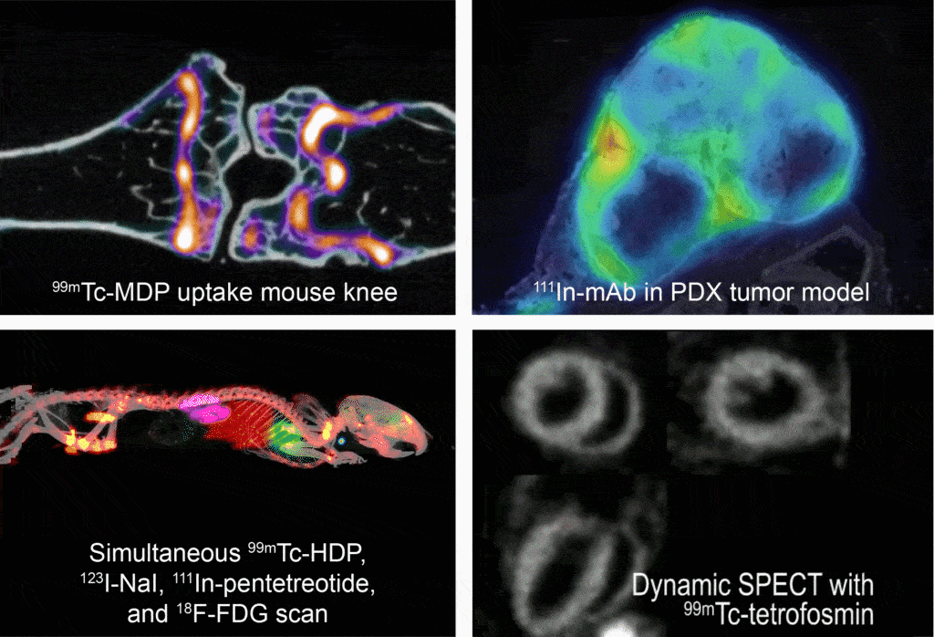
Image Credit: MiLabs - Molecular Imaging Solutions
The multimodal approach of U-SPECT7 with structural/anatomical images obtained with our high-speed CT scanner, namely U-CT, has been increasingly accepted in the preclinical arena. The combination of SPECT/CT presents added and more accurate diagnostic information compared to either in isolation. Another benefit of this configuration is that it enhances reconstruction and improves quantification utilizing the combined information from these two imaging modalities. The modular nature of the system means you can upgrade a U-SPECT7 to U-SPECT7CT with CT capabilities at a later date to suit your technical or financial requirements.
Applications
- Theranostics - U-SPECT can image at sub-mm resolutions photon energies up to 1 MeV. This makes direct imaging and quantitation of targeted theranostics (radiotherapies) possible for a wide range of high-energy therapeutic isotopes including 131I, and alpha particle emitters such as 212Pb, 225Ac, 213Bi, 221Fr, 209At/211At, 223Ra and many others.
- Quantitative 3D Auto-radiography - By scanning cryo-cooled tissue samples, such as complete organs, with a dedicated super-resolution SPECT collimator, researchers can obtain 3D auto-radiography distributions at 0.12 mm resolution.
- Oncology
- Neurology
- Immunology
- Cardiology
- Infectiology - including COVID-19 related studies
- and many more